Le Maître A, Guy F, Merceron G et al. 2022
Int J Primatol (2022). doi.org/10.1007/s10764-022-00329-4
Published online 20 September 2022
News article at the Faculty of Lifesciences, University of Vienna
Abstract
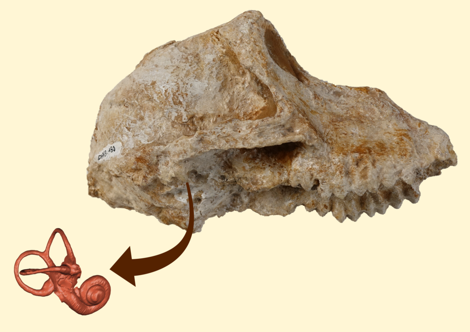
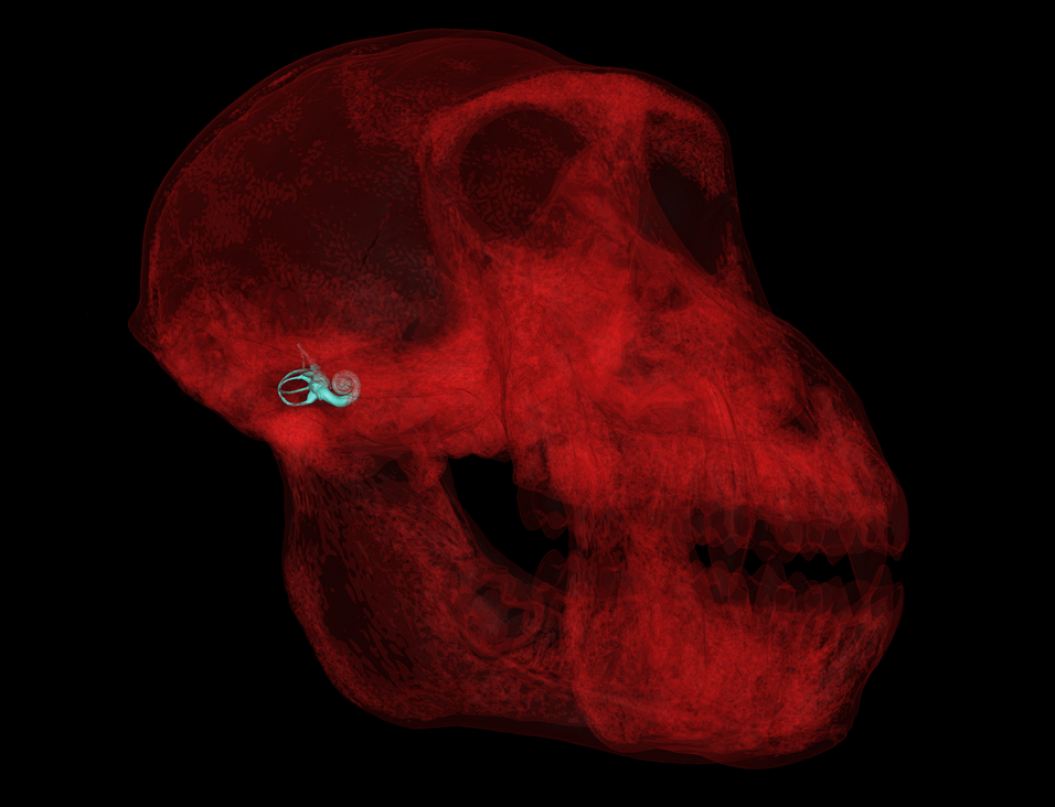
Discoveries in recent decades indicate that the large papionin monkeys Paradolipopithecus and Procynocephalus are key members of the Late Pliocene – Early Pleistocene mammalian faunas of Eurasia. However, their taxonomical status, phylogenetic relationships, and ecological profile remain unclear. Here we investigate the two latter aspects through the study of the inner ear anatomy, as revealed by applying micro-CT scan imaging techniques on the cranium LGPUT DFN3-150 of Paradolichopithecus from the lower Pleistocene (2.3 Ma) fossil site Dafnero-3 in Northwestern Greece. Using geometric morphometric methods, we quantified shape variation and the allometric and phylogenetic signals in extant cercopithecines (n = 80), and explored the morphological affinities of the fossil specimen with extant taxa. LGPUT DFN3-150 has a large centroid size similar to that of baboons and their relatives. It shares several shape features with Macacina and Cercopithecini, which we interpret as probable retention of a primitive morphology. Overall, its inner ear morphology is more consistent with a stem Papionini more closely related to Papionina than Macacina, or to a basal crown Papionina. Our results, along with morphometrical and ecological features from previous studies, call into question the traditional hypothesis of a Paradolichopithecus-Macacina clade, and provide alternative perspectives in the study of Eurasian primate evolution during the late Neogene-Quaternary.