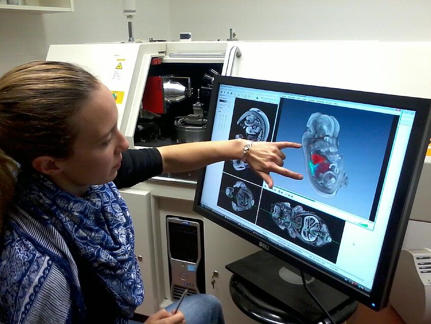
The MicroCT Imaging Lab
X-ray microtomography (microCT) provides non-destructive detailed 3D imaging of a wide variety of sample types, from contrast-enhanced embryos and tissue specimens to dried insects and small fossils. We apply microCT to numerous research problems in development and evolution, including comparative embryology, functional morphology, and morphometric studies. We are not a service facility, but we are interested in discussing productive collaborations.
© Brian Metscher
The lab has three microCT scanning systems, all self-contained (i.e. no synchrotron source and individually shielded):
- a Zeiss Xradia MicroXCT system capable of tomographic imaging of biological objects from about 12mm down to less than 500µm in size, with possible resolutions below 2µm;
- a Bruker SkyScan 1174 for lower resolution imaging of larger specimens. This is a low-cost scanner made for samples up to about 30mm, and gives spatial resolutions down to about 20µm;
- a new Bruker SkyScan 1272 for diverse specimens of moderate density from 5-10mm to a few cm in size. This system will begin service in mid-2021.
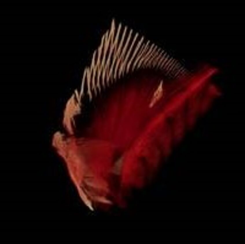
MicroCT images
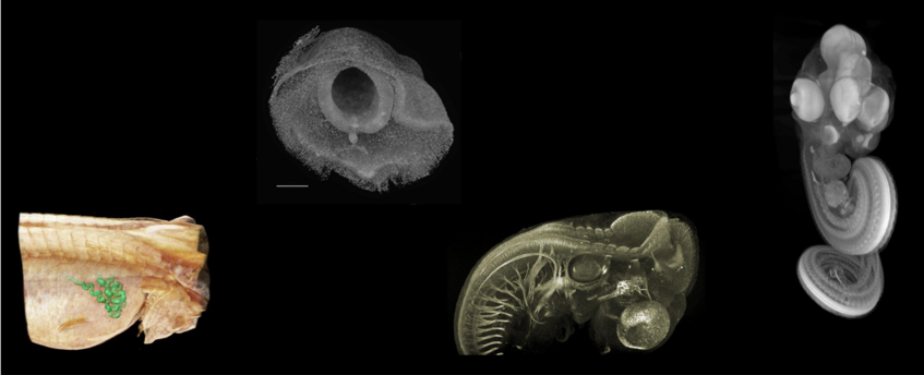
© Brian Metscher
Videos
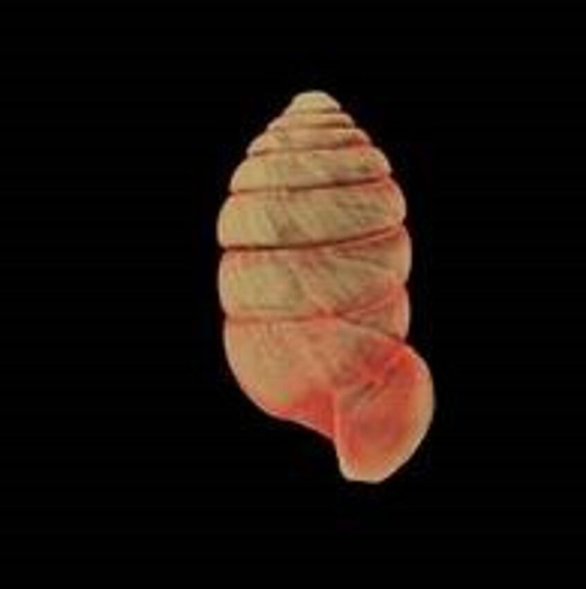
Orcula
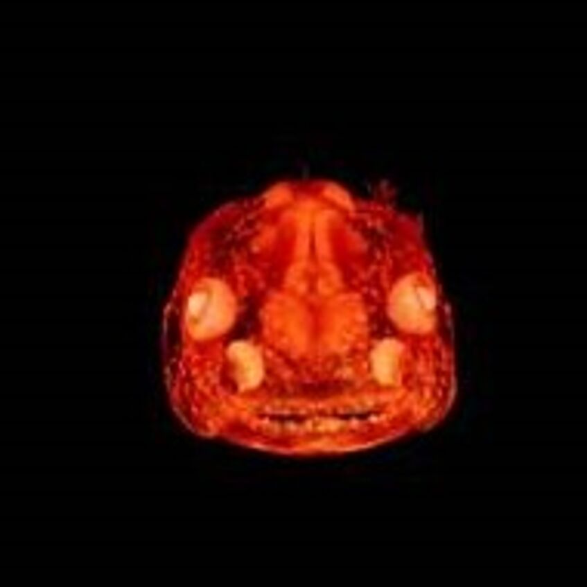
Axolotl
Sturgeon
Selected publications
Akkari, N., Ganske, A. S., Komerički, A., & Metscher, B. (2018). New avatars for Myriapods: Complete 3D morphology of type specimens transcends conventional species description (Myriapoda, Chilopoda). PLOS ONE, 13(7), e0200158. doi.org/10.1371/journal.pone.0200158
Handschuh, S., Beisser, C., Ruthensteiner, B., & Metscher, B. (2017). Microscopic dual‐energy CT (microDECT): a flexible tool for multichannel ex vivo 3D imaging of biological specimens. Journal of Microscopy, 267, 3-26. doi.org/10.1111/jmi.12543
Akkari, N., Enghoff, H., & Metscher, B. D. (2015). A New Dimension in Documenting New Species: High-Detail Imaging for Myriapod Taxonomy and First 3D Cybertype of a New Millipede Species (Diplopoda, Julida, Julidae). PLOS ONE, 10(8), e0135243. doi.org/10.1371/journal.pone.0135243
Metscher, B. D., & Müller, G. B. (2011). MicroCT for molecular imaging: Quantitative visualization of complete three‐dimensional distributions of gene products in embryonic limbs. Developmental Dynamics, 240, 2301-2308. doi.org/10.1002/dvdy.22733
Metscher, B. D. (2009). MicroCT for comparative morphology: simple staining methods allow high-contrast 3D imaging of diverse non-mineralized animal tissues. BMC Physiology, 9, 11. doi.org/10.1186/1472-6793-9-11
Metscher, B. D. (2009). MicroCT for developmental biology: A versatile tool for high‐contrast 3D imaging at histological resolutions. Developmental Dynamics, 238, 632-640. doi.org/10.1002/dvdy.21857